

A Timeline of Our Profession
In November 1895, Wilhelm Conrad Roentgen discovered mysterious rays that could pass through most substances, casting shadows of solid objects. He named them X-rays, after the algebraic term for an unknown quantity. Soon, medical practitioners were using X-rays to identify bone structures, locate foreign objects in the body, and perform other types of medical imaging.
A year later, Antoine Henri Becquerel began to study radioactivity and look for natural sources of radiation. Marie Curie and Pierre Curie in 1898 discovered two radioactive elements: radium and polonium. By 1901, doctors were testing radium on skin lesions and using it to treat lupus and cancer at the Saint-Louis Hospital in Paris. Roentgen and the Curies would later win Nobel Prizes in Physics.
Their discoveries led to the job of X-ray technician—and now to the profession of radiologic technologist. Today’s technologists work throughout health care, performing medical imaging, interventional procedures, and radiation therapy. Follow our timeline to see highlights from the profession’s history.
1896
Enrico Salvioni invents, Thomas Edison improves the first commercial fluoroscope to take radiographs (X-rays).
1904
Following the first deaths from exposure to radiation, practitioners begin covering themselves and their patients in lead shields, aprons, and gloves.
1913
William Coolidge invents the hot cathode X-ray tube, which is more dependable than previous versions and can treat deeper cancers.
1914
Marie Curie invents a mobile X-ray unit, enabling medics to scan wounded soldiers near battlefields during World War I.
1917
The U.S. Army Medical Corps establishes intensive programs to train medical officers as radiologists and enlisted men as technicians.
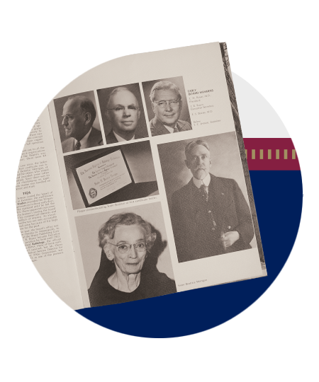
1922
The Radiological Society of North America (RSNA)—with support from the American Roentgen Ray Society and the American Society of X-Ray Technicians (now ASRT)—founds what is now ARRT.
1922
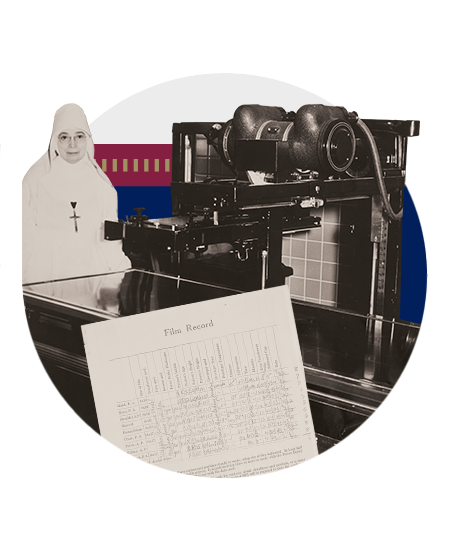
ARRT administers our first Radiography exam to Sister Mary Beatrice Merrigan. After answering 20 essay questions and submitting 10 required radiographic films, she becomes ARRT’s first R.T.
1928
The Second International Congress of Radiology defines an international unit of radiation exposure—the roentgen—which enables physicists to reliably compare doses and results.
1936
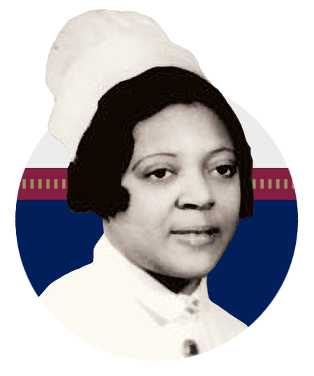
Rose Marie Pegues, R.N., becomes the first Black R.T.1
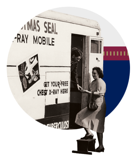
1940s
Radiographers conduct chest X-rays in schools, workplaces, and clinics, screening for tuberculosis before patients become seriously ill. 2
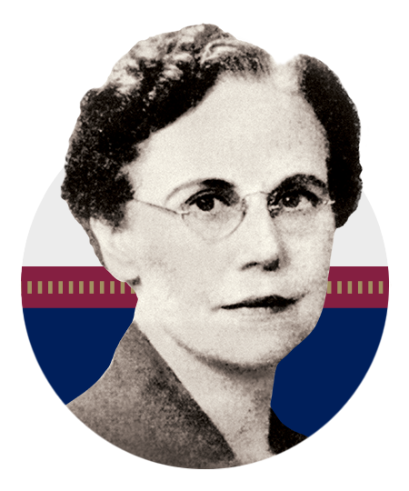
1943
Erminda R. Clarke, R.T., of Lincoln, Nebraska, becomes the first woman to serve on our Board of Trustees. 3
1945
Tests and deployment of atomic bombs help bring an end to World War II, broaden awareness of the effects of radiation, and lead to the use of atomic energy in nuclear medicine.
1947
Sister Mary Alacoque Anger is the first person to receive a bachelor’s degree in radiologic technology.

1954
The ARRT exam eliminates sample X-rays. The next year, ARRT drops the essay component and moves to all multiple-choice questions.
1958
U.S. cardiologist F. Mason Sones Jr. mistakenly injects the small vessels of a patient’s heart with a significant amount of contrast dye. The error ultimately leads to modern cardiac imaging.
1959
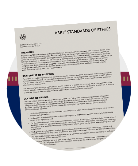
ARRT publishes its first Code of Ethics
1960
The world’s first peacetime hospital ship—the S.S. Hope—launches. Many R.T.s will serve on the ship, which brings U.S. health care techniques to developing nations.
1962
ARRT adopts the more inclusive term “radiologic technologist” over “X-ray technician.”
1963
The first U.S. cyclotron begins operation at Washington University Medical School. By manufacturing radioisotopes, it reduces the need for natural radioactive sources (such as radium) by manufacturing radioisotopes.
1963
First ARRT Nuclear Medicine Technology exam
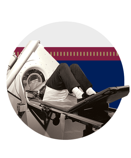
1967
Godfrey Hounsfield invents the CT scanner, which increases by 100 times the amount of information in each image. 4
1969
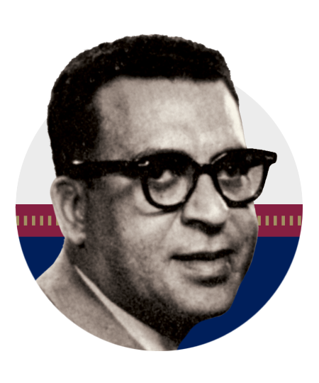
Royce Osborn, R.T., becomes the first Black president of ASRT. Today, ARRT funds a scholarship program that honors him. 5
1973
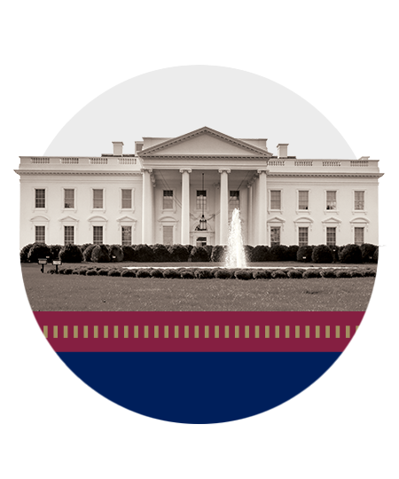
To commemorate ARRT’s 50th anniversary, First Lady Patricia Nixon—a former radiographer—invites organizational representatives to the White House for tea.
1973
Mayo Clinic installs the first CT scanner in the U.S.
1976
The American Cancer Society recommends mammography as a method of screening for breast cancer.
1977
Raymond Damadian, M.D., along with Lawrence Minkoff and Michael Goldsmith, perform the first MRI body scan of a human being.
1983
Nuclear medicine specialist Henry Wagner Jr., M.D., uses a positron emission tomography (PET) scanner to take an image of a radioactive tracer in his own brain.
1986
Ultrasound technology improves, resulting in the first 3D image of a fetus. By the late 1990s, 4D ultrasounds can show movement in real time.
1990
ARRT adopts its Standards of Ethics.
1991
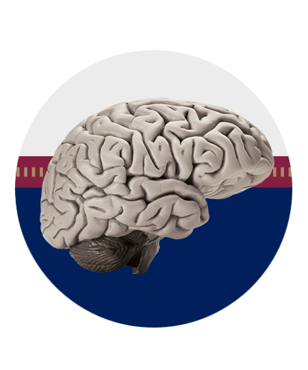
John Belliveau presents images of human brain activity using functional MRI, a process that measures changes in blood flow that correspond with brain activity.
1991
First ARRT Cardiovascular Interventional Radiography* and Mammography exams

1991
ARRT administers our first “advanced level” (now postprimary) exams.
1995
First ARRT MRI and CT exams
1995
ARRT adopts biennial continuing education requirements to help ensure that R.T.s stay up to date with their knowledge.
1995
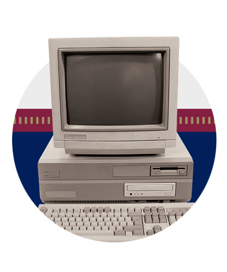
DuPont Diagnostic Imaging introduces a system that converts X-rays into electronic data, making it possible to immediately view images on a screen instead of having to develop film.
1997
First ARRT Quality Management* exam
1999
ARRT begins the transition to computer-based exams, enabling candidates to take an exam throughout the year at locations across the U.S.
1999
30% of R.T.s held more than one ARRT credential, up from 5% in 1991. Today, in 2022, it’s nearly 47%.
2000
First ARRT Sonography exam
2001
Nearly 17 million nuclear medicine procedures take place in the U.S. this year.
2001
First ARRT Vascular Sonography and Bone Densitometry exams
2003
ARRT splits Cardiovascular Interventional Radiography exam and administers our first Cardiac Interventional Radiography and Vascular Interventional Radiography exams
2004
First ARRT Breast Sonography exam
2005
ARRT launches a certification process for a new role, the Registered Radiologist Assistant (R.R.A.)
2007
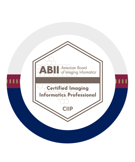
ARRT and the Society for Imaging Informatics in Medicine form the American Board of Imaging Informatics (ABII), which offers certification to imaging informatics professionals.
2008
A new generation CT scanner makes it possible to take images of the heart and coronary arteries in less than one second.
2015
The ASRT Museum and Archives opens in Albuquerque, New Mexico. Its artifacts and exhibits tell the history of the radiologic technology profession.
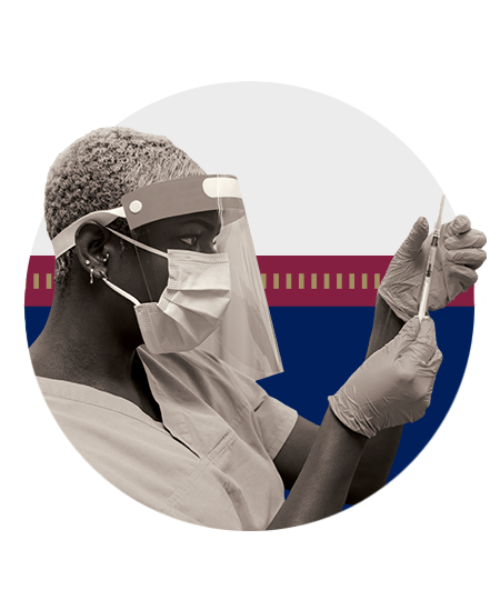
2020
The worldwide COVID-19 pandemic severely disrupts every part of society including health care as a whole and technologist education programs.
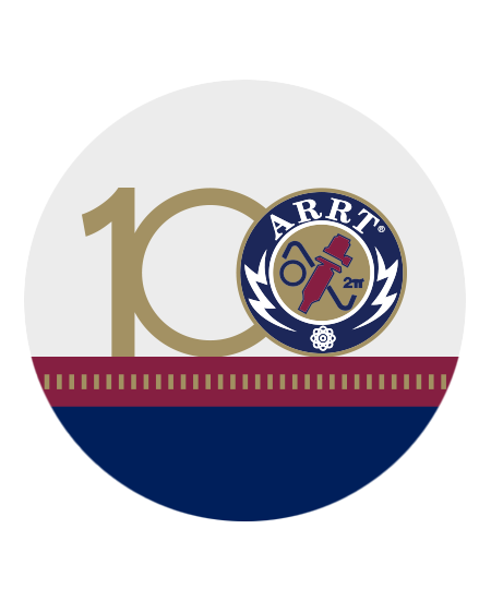
2022
ARRT and R.T.s celebrate a century of gold standard patient care.
1: The Shadowmakers: A History of Radiologic Technology, American Society of Radiologic Technologists, 2020, (Page 45)
2: The Shadowmakers: A History of Radiologic Technology, American Society of Radiologic Technologists, 2020, (Cover 2)
3: Image courtesy of American Society of Radiologic Technologists
4: The Shadowmakers: A History of Radiologic Technology, American Society of Radiologic Technologists, 2020, (Page 224)
5: The Shadowmakers: A History of Radiologic Technology, American Society of Radiologic Technologists, 2020, (Page 82)
* We no longer issue new credentials for Quality Management and Cardiovascular Interventional Radiography. People who hold these credentials can maintain them indefinitely, however, if they continue to meet our ethical standards and other requirements for doing so.